Events
Nov 25, 2025
Seminar by Dr. Moe Mahjoub; Talk title: Seeing is believing: Discovery of a new structure essential for multiciliogenesis
School of Biomedical Sciences cordially invites you to join the following seminar:
Speaker: Dr. Moe Mahjoub, Associate Professor, Departments of Medicine (Nephrology), Cell Biology and Physiology, Washington University in St. Louis
Talk Title: Seeing is believing: Discovery of a new structure essential for multiciliogenesis
Date: 25 November 2025 (Tuesday)
Time: 3:30 pm – 4:30 pm
Venue: Seminar Room 2, 4/F, 3 Sassoon Road
Host: Professor Mu He
Biography
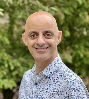
Dr. Mahjoub received his Ph.D. in Molecular Biology and Biochemistry from Simon Fraser University in Canada, and completed postdoctoral training at Stanford University. He is an Associate Professor (with tenure) in the Departments of Medicine, Cell Biology & Physiology at Washington University in St. Louis. His laboratory investigates the role of the cell cytoskeleton in regulating tissue formation, focusing on microtubule-based organelles called centrosomes and cilia. They use innovative tools to define the critical functions of these organelles during mammalian organ development, using a combination of transgenic mouse models, in vitro organoid cultures, and advanced microscopy techniques. Dr. Mahjoub serves on the Science Advisory Panel of the Polycystic Kidney Disease Foundation, as well as several national and international grant review panels. He is also Co-Director of the WashU Ciliopathy Research Group, a multidisciplinary team of investigators that perform fundamental, translational, and clinical research on human ciliopathies.
Abstract
Centriolar satellites are specialized non-membranous granules that act as conduits for the transport and degradation of proteins in the cytoplasm. Yet, our understanding of satellite structure, function, and dynamics in different tissues in vivo remains limited. To address this, we developed a “SatelliteGFP” reporter mouse model that expresses a GFP-tagged PCM1 fusion protein under its native promoter in all cell types. PCM1 is the key scaffolding protein essential for the assembly and function of satellite granules. Immunofluorescence analysis of organs isolated from PCM1GFP/GFP mice showed dramatic variations in the expression levels, size, and distribution of PCM1-positive condensates in different ciliated cells. Comparative proteomics identified organ-specific differences in the PCM1-satellite interactome, further highlighting their compositional heterogeneity in vivo. Intriguingly, we discovered a novel PCM1-associated structure in multiciliated cells which we have named the Fibrogranular Material Reservoir (FGM-R). Using extended live-cell imaging, we defined the kinetics of FGM-R biogenesis, movement, and dissolution during multiciliated cell differentiation. Our results shed light on the heterogeneity of satellite granule dynamics and composition in ciliated cell types impacted in ciliopathy patients.
ALL ARE WELCOME.